|
Lääketieteellisen fysiikan ja tekniikan yhdistys (LFTY) Finnish Society for Medical Physics and Medical Engineering |


X Finnish Medical Physics and Medical Engineering Day, 10.2.2011, Aalto University
The tenth Finnish Medical Physics and Medical Engineering Day was held 10 February 2011 at Aalto University, Otaniemi, Espoo, Finland. The annual event gathered this year 98 student and researcher participants and ten Finnish companies from the area of medical physics and medical engineering. The traditional poster exhibition, where the best Master's theses and diploma theses finished in 2010 were awarded had 11 participants. The award for the best theses was this year 2010 €.From the 11 participants, one thesis was reckoned to be above the others. This thesis was:
- Harri Kokkonen, University of Eastern Finland
Detection of Mechanical Injury of Articular Cartilage Using Contrast Enhanced Computed Tomography
- Nikolai Beev, Tampere University of Technology
Assessment of Sternal Instability by Vibration - Aleksi Iho, University of Helsinki/Aalto University
Targeting cancer cells: a molecular engineering approach - Paavo Vartiainen, University of Eastern Finland
Using Kalman filter and hierarchical model in the analysis of motion laboratory measurement data
 |
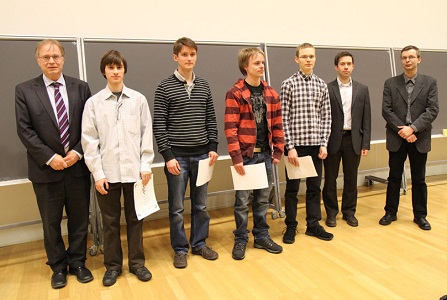
| Award presentation for the best Master's theses and diploma work in 2010 (from left to right): Prof. Risto Ilmoniemi (chairman of Finnish Medical Physics and Medical Engineering Society), Nikolai Beev, Aleksi Iho, Harri Kokkonen, Paavo Vartiainen, Samu Taulu (Elekta Oy), and Mika Tarvainen (secretary of Finnish Medical Physics and Medical Engineering Society).. |
Abstracts of the awarded theses
Detection of Mechanical Injury of Articular Cartilage Using Contrast Enhanced Computed Tomography
Harri Kokkonen, University of Eastern Finland
Objective: Osteoarthritic degeneration may be initiated by mechanical overloading of articular cartilage. Mechanical injury increases the permeability of tissue, thereby probably affecting the diffusion of contrast agents in articular cartilage. We investigated whether it is possible to detect acute cartilage injury by measuring contrast agent diffusion into articular cartilage using contrast enhanced computed tomography (CECT).
Methods: Osteochondral plugs (Ø=6.0mm, n=36) were prepared from intact bovine patellae (n=9). Two of the adjacent samples were injured by impact loading, using a drop tower, while the others served as paired controls. The samples were imaged before immersion in contrast agent solution (ioxaglate (Hexabrix™) or sodium iodide (NaI)) and 1, 3, 5, 7, 10, 15, 20 and 25 hours after immersion using a MicroCT-instrument. Contrast agent content, diffusion coefficient and diffusion flux were determined for each sample.
Results: Already after one hour the penetration of contrast agents into cartilage was significantly (p<0.05) greater in the injured samples. The diffusion coefficient was not altered by the injury, which suggests that reaching the diffusion equilibrium takes the same time in injured and intact cartilage. However, the diffusion flux of ioxaglate through the articular surface was significantly higher in injured samples at 30 to 60 minutes after immersion.
Conclusions: To conclude, CECT could diagnose articular cartilage injuries, and determination of the diffusion flux of ioxaglate helped to detect tissue injury without waiting for the diffusion equilibrium. These results are encouraging, however, in vivo application of CECT is challenging and systematic further studies are needed to reveal its clinical potential.
Assessment of Sternal Instability by Vibration
Nikolai Beev, Tampere University of Technology
Objective: The purpose of the study conducted at Tampere University of Technology and Tampere University Hospital was to devise a novel method for detection of instability or abnormal healing in the sternum after open chest surgery. Patients suffering from such postoperative complications are exposed to higher risk of deep wound infection and other severe complications. The proposed method, based on vibration measurement, aims to provide quantitative information on the mechanical stability of the breastbone that cannot be obtained from clinical examination or medical imaging techniques.
Methods and materials: A special measurement system was designed and constructed at the Tampere University of Technology. The system produced controlled vibration with spectral content in the 50-1500 Hz range. The response of the studied breastbone was picked up by an accelerometer and recorded in digital format. The collected data was processed on a personal computer using routines implemented in MATLAB. The mechanical transfer function of the sternum was estimated and several descriptive parameters were extracted from it. Their distributions were studied to determine how they reflect changes occurring in the breastbone.
Altogether 22 elective cardiac surgery patients from the Pirkanmaa district were recruited for the study. Complete datasets are available for 12 patients with uneventful healing. Five patients were diagnosed with sternal instability. Measurements were taken at 3 levels of the sternum: upper, middle and lower. Four measurement sessions were organized for each patient: preoperative, early postoperative (typically 3rd-4th postoperative day), 3rd postoperative week and 3rd postoperative month. In addition to vibration stimulation, manual palpation was applied to provoke movement of the sternal halves and the resulting acoustic emissions in case of instability were recorded and studied.
Results: The middle and lower measurement positions were found to be consistent and yielding useful data, unlike the upper position. The most important changes occur in the higher frequency (600-1500 Hz) band of the studied transfer functions and are reflected by derived parameters such as the integrated transmittance in this band. Significant changes caused by the introduced median incision and subsequent healing processes are observed: shortly after the operation vibration transmittance in the 600-1500 Hz band drops significantly, after which it increases gradually. Measured postoperative values higher than the preoperative suggest auxiliary transmission of vibration, possibly due to the introduced steel wire fixations. The statistical significance of changes in parameter distributions was confirmed by the Wilcoxon signed-rank test. Artifacts produced by provoked instability are clearly identifiable in the time-frequency representation of recorded signals.
Conclusions: The proposed method was implemented using specially built instrumentation and studied in clinical setting. Results confirmed that mechanical changes in the sternal incision are reflected in the vibration response. It was also demonstrated that acoustic emissions caused by provoked instability could be identified. Vibration measurements are painless, safe and do not expose the patient to ionizing radiation. More consistent measurement data from subjects with sternal instability is needed to further develop the technique.
Targeting cancer cells: a molecular engineering approach
Aleksi Iho, University of Helsinki/Aalto University
The cellular membrane is a highly complex mixture of lipid molecules and proteins with each molecule playing specific part in the functional role of the cell. Phosphatidylserine (PS) is an anionic lipid, that accounts for up to 20% of the total lipids in a eukaryote cell membrane. The lipid is normally located almost exclusively in the inner leaflet of the lipid bilayer. However, cancerous cells are found to express elevated levels of PS on the outer leaflet of the plasma membrane.
In this study a short peptide (18 residues) was designed with the aim that it could be used as a chemical reporter to target the expression of PS on the outer leaflet of the plasma membrane in cancer cells. The peptide would combine PS binding properties as well as antimicrobial peptide (AMP) -like functionality that would enable it to penetrate and anchor itself to the membrane. The design involves a two-phase targeting strategy, in which click-chemistry reactions are used to introduce imaging moiety to the peptide after the peptide has found the target.
A peptide was designed by modeling random sequences on software available online, and looking for AMP-like structural elements. The sequence was combined with a segment found in literature. A series of biophysical experiments, including peptide intercalation into lipid monolayers and Förster resonance energy transfer (FRET) experiment, were then conducted to study the peptide's properties and to confirm the utility of the two-phase labeling strategy. A cell labeling experiment was carried out with cancerous HeLa cells and healthy mouse fibroblast (3T3) cells to test the peptide’s selectivity with live cells.
The peptide showed significant specificity towards anionic PS and desired AMP-like characteristics in the presence of PS with model membranes. The two-phase labeling strategy was found to be functional. The cell labeling results were promising, showing a degree of selective labeling of HeLa cells, but the results cannot be considered conclusive yet due to some limitations in the experiment.
This study has shown that a molecular engineering approach into designing functional biomolecules is feasible. A peptide was designed from modeling principles and was evaluated to have several of the predesigned, desired features. The results support the hypothesis that PS exposed in cancer cells could be used as a molecular target for sensitive diagnostic imaging of tumors and metastases. Only a single peptide was tested in the present study, and there is reason to believe the design can be iterated to enhance the functionality of the peptide significantly. The future designs of functional peptides will have great potential in various research and clinical applications.
Using Kalman filter and hierarchical model in the analysis of motion laboratory measurement data
Paavo Vartiainen, University of Eastern Finland
Motion analysis is study of human motion, for example gait. Motion analysis is used to study neurological disorders and musculo-skeletal disorders. Motion analysis is also used in entertainment industry and in ergonomics research.
The aim of this study was to model human gait and to estimate force between sole and the floor using only data recorded with camera system.
In motion laboratory the motion of the subject is recorded using reflecting balls attached on the anatomical points on subject’s skin. These balls are imaged with multiple cameras and thus the trajectory of the balls can be reconstructed.
In the study human motion was modeled by using so called hierarchical body model and Kalman filter and Kalman smoother algorithms. Body model consists of rigid objects joined together by joints. Hierarchical means that the pose of the model in 3d-space is defined by location of one object and by joint angles.
Model can be used eg. to study motion of hip, knee and ankle joints. To formulate observation model for Kalman filter points that correspond the locations of reflecting balls are fixed on the model objects.
Kalman filter and kalman smoother are used to estimate pose, velocities and accelerations of the body model. By using acceleration estimates and masses of the body segments one can estimate forces. In this study the force between soles and the floor was estimated. The estimated force was compared to the measured force. Estimation of the vertical component of this reaction force was fairly accurate.
The modeling of gait with methods presented in the work is possible even when some of the observations are temporary missing; which is often the case with motion laboratory data. Kalman filter can be tuned so that missing and inaccurate observations do not affect to the estimates. Furthermore Kalman filter has wide tuning possibilities. By tuning filter parameters one can effectively diminish the effect of measurement noise to the estimates.