|
Lääketieteellisen fysiikan ja tekniikan yhdistys (LFTY) Finnish Society for Medical Physics and Medical Engineering |

VIII Finnish Medical Physics and Medical Engineering Day, 12.2.2009, University of Kuopio
The eight Finnish Medical Physics and Medical Engineering Day was held 12 February 2009 at University of Kuopio, Kuopio, Finland. The annual event gathered this year 90 student and researcher participants and seven Finnish companies from the area of medical physics and medical engineering. The traditional poster exhibition, where the best Master's theses and diploma theses finished in 2008 were awarded had 8 participants. The award for the best theses was this year 2008 &euro.From the 8 participants, two theses were reckoned to be above the others. These two theses were:
- Sari Ahokas, Tampere University of Technology
Development of low noise active electrodes for EEG measurements - Jaakko Nieminen, Helsinki University of Technology
Enhancement of MRI with SQUID arrays and by polarization encoding
- Timo Vuorela, Tampere University of Technology
Unraveling lipoprotein structure via molecular dynamics
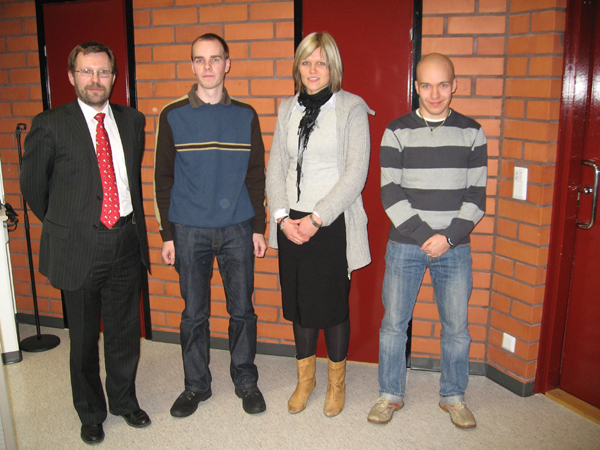 |
| Award presentation for the best Master's theses and diploma work in 2008 (from left to right): Prof. Pasi Karjalainen (chairman of Finnish Medical Physics and Medical Engineering Society), Jaakko Nieminen, Sari Ahokas and Timo Vuorela. |
Abstracts of the awarded theses
Development of low noise active electrodes for EEG measurements
Ahokas Sari, Tampereen teknillinen yliopisto
Nowadays many brain investigation methods exist. EEG measures the electrical activity of the brain and magnetoencephalography (MEG) detects the magnetic fields that are a result of the electrical activity. Computed tomography (CT) describes the brain anatomy. Functional Magnetic Resonance Imaging (fMRI) detects the changes in the blood flow after nerve cell activation. Spectral Emission Tomography (SPECT) and Positron Emission Tomography (PET) measure the metabolic activity of the brain. The advantage of EEG and MEG over the other brain investigation methods is their high temporal resolution and therefore the possibility to study the brain function in millisecond time scale. Their disadvantage is a poor spatial resolution that limits the possibility to find an exact source location. It was long believed that MEG offers better spatial resolution than EEG due to the high skull resistivity. Nowadays it has been proven that the skull resistivity is not as high as was thought and that EEG actually offers at least as good spatial resolution as MEG or even better. The EEG recording system is cheaper and smaller than the MEG device and it has lower restrictions for the patient movement. In the EEG measurement the spatial resolution can be increased by using larger number of electrodes and low noise measurement instruments. The advantages of EEG measurement and possibilities of better signal quality make it an interesting research topic.
Nowadays due to the development of electronics and especially integrated circuits the possibilities to design small sized active electrodes exist. Active electrodes are a developed version of commonly known passive electrodes and they have an electronic circuit integrated into the electrode. Active electrodes act as an impedance converter and can also have other properties such as amplification, filtering or impedance detection. Active electrodes are more noise tolerant and do not require skin preparation. The need for active electrodes exists especially in high resolution EEG measurements were large number of electrodes are used to obtain the best possible accuracy and signal quality, and in emergency medicine where fast electrode placement is essential. This Master of Science thesis is part of an ongoing research project where objectives are to develop methods and instrumentation to record electric fields of the brain more accurately. The main purpose of this project was to develop a simple and low noise active electrode that can be used in high resolution EEG measurements. Also solutions for electrode wiring were discussed during the design process.
Enhancement of MRI with SQUID arrays and by polarization encoding
Nieminen Jaakko, Teknillinen korkeakoulu
Magnetic resonance imaging (MRI) is a method to study the interior structure of matter. It is based on detecting precession signals from a magnetized sample. The detection can be speeded up by using sensor arrays. In low-field MRI, a sample is polarized in a magnetic field of several millitesla. The polarization is followed by SQUID-based signal detection at microtesla-region fields. In this study, a new encoding method to reduce imaging times in low-field MRI is developed.
In MRI, signals of a sensor array can be written in vector form s(t) = Am(t), where A is a lead field matrix and m(t) contains the components of the voxel magnetizations. In polarization encoding, various polarizing fields affect the initial magnetization of the sample in consecutive measurements. Assume that for the kth measurement the magnetizations are m_k(t) = C_km(t), where C_k is a conversion matrix. Then s_k(t) = AC_km(t). A large signal vector s and a generalized lead field matrix A are constructed combining s_k:s and AC_k:s row-wise, respectively. Then, s(t) = Am(t). If the polarizing fields are chosen properly, rank A > rank A, i.e., the number of linearly independent sensors is increased.
The proposed method was tested with low-field MRI simulations using the geometry of a 304 channel SQUID system. Noisy signals from a 15×15 voxels phantom were simulated to the SQUIDs. The inverse problem of image reconstruction was solved using truncated singular value decomposition. The results show that by increasing the number of polarizing fields, the imaging times are reduced. Equally, in a given time the imaging quality can be improved by increasing the number of polarizing fields.
In this study, a new encoding method was developed for MRI. Polarization encoding introduces additional independent sensors and increases the information about the sample. The method reduces imaging times. It is possible to combine the method with other encoding methods; for instance, one dimension can be encoded by polarization encoding and the others by Fourier encoding techniques. Polarization encoding is especially suitable for low-field MRI, because at low fields it is possible to construct sets of polarizing fields. Moreover, at low fields, imaging processes need improvements because current methods are slow and suffer from poor image quality. In addition to MRI, polarization encoding can also be used to improve other imaging techniques, such as magnetorelaxometry.
Unraveling lipoprotein structure via molecular dynamics
Vuorela Timo, Tampereen teknillinen yliopisto
Cardiovascular diseases are the primary cause of death in the industrialized countries. Heart strokes and cerebral infarctions are the end conditions of a cardiovascular disease called atherosclerosis. According to the current view, it is inflicted and further restrained by lipoproteins circulating in blood. In particular, low density lipoprotein (LDL) levels have been found to correlate positively and high density lipoprotein (HDL) levels inversely with the risk of atherosclerosis.
Lipoproteins are complex macromolecular structures designed to carry cholesterol in an esterified form inside the body. They consist of a spherical lipid part surrounded partly by a protein part. Although molecular compositions are quite well understood, the molecular-scale structures have not been solved. In this study we use new computational methods to get information about the molecular-scale ordering in different sized lipoprotein particles.
In this study the structure of two different lipoprotein particles were explored by using classical molecular dynamics methods. The smaller system was constructed to model the lipid part of an HDL particle and the larger system the lipid part of an LDL particle. The LDL particle contains over 3000 molecules and is more than three times larger than the systems in current state-of-the-art studies. Also the time scales in this study are over a magnitude longer than in the earlier studies. To achieve the length and time scales used in this study a method called coarse graining has been used. In coarse graining on average four carbon atoms are substituted by one bead. Coarse graining reduces the complexity of the systems while preserving the behavior of different molecules. Extensive structural analysis is performed for the general structure as well as conformations of each molecule type. Also the dynamics of the whole lipoprotein particles as well as the individual components is studied.
The current view of the lipoprotein structure is that the particles are composed of a disordered hydrophobic core and a hydrophilic surface. The results of this study support the view, but also reveal that there is an additional layer between the core and the surface. The results show significant ordering of the core lipids close to the surface that is not observed in the core region. Cholesteryl esther molecules in the interface order parallel to the phospholipid tails similar to cholesterol molecules. Also triacylglycerols in the interface have more compact packing and have a tendency to orientate in a way that minimizes contacts with water. The diffusion rates of different components are in good agreement with the results from earlier experimental and computational studies.
This study demonstrates that molecular dynamics and especially coarse grained methods can be used to give valuable insight into the structures of lipoproteins, unreachable to other methods. The results are in line with the current view of the lipoprotein particle structure and, in addition, support a recently presented new three-layer structure for lipoprotein particles. This study gives a solid foundation for future projects which aim to better understanding about the structure-function relationship of lipoproteins.